The coronascope on television

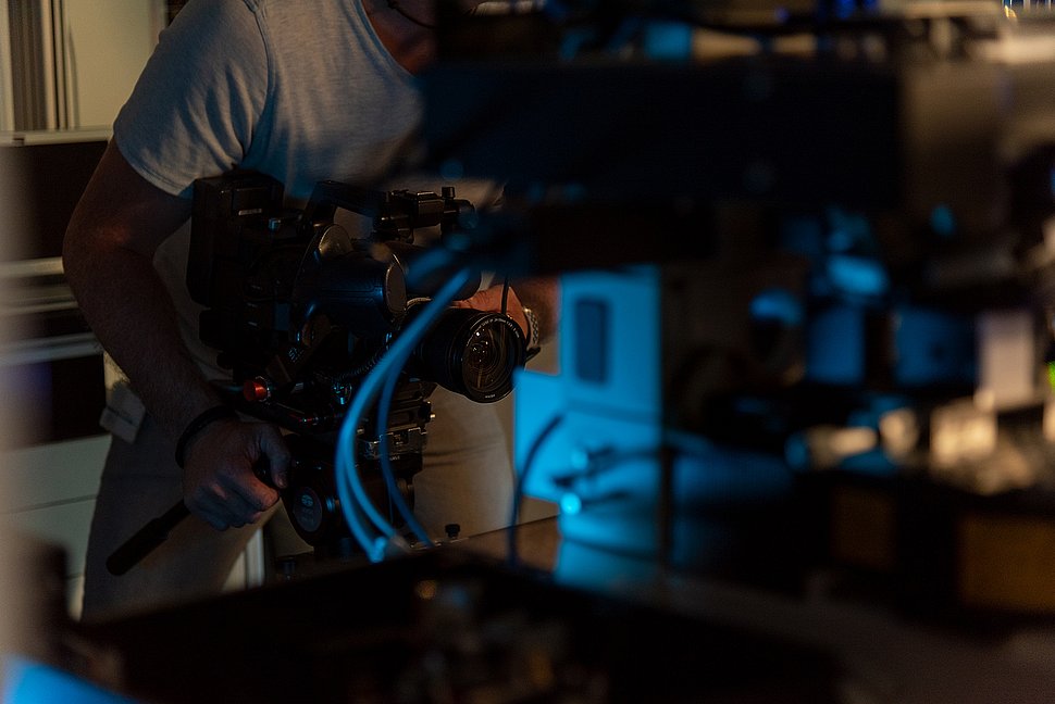
Recently, a film team of the Bayerischer Rundfunk (Bavarian broadcasting organisation) visited the Max Planck Institute for the Science of Light: The programme about the coronascope, which is currently located in the virus laboratory of the University Hospital Erlangen and takes pictures of cells infected with the Sars-CoV-2 virus, was broadcasted yesterday on 3sat in the science programme nano. Click here for the video clip (German).
For all non-German-speaker, here is a transcript of the video:
Will we soon be able to use laser light to better understand the corona virus?
Scientists at the Max Planck Institute in Erlangen are pursuing this idea. A new light microscope developed here can show the inner life of cells with enormous accuracy. The researchers hope that it will soon be possible to observe the complete reproduction cycle of the virus in the cell as a film.
Vahid Sandoghdar: Light microscopy has the advantage that I can examine living cells, i.e. I can see in real time what the cell is doing and in our case hopefully what the virus is doing.
But here at the Institute of the Science of Light, the scientists cannot work with the dangerous corona virus. The investigations are therefore carried out a few kilometres away in a high-security laboratory of the Virological Institute at the University Hospital Erlangen. This is where the first prototype of the coronascope is located - in the safety level 3 laboratory, which may only be entered by a few specialists.
But why do scientists rely on light microscopes at all when researching the virus? After all, this technology seems somewhat outdated at first glance. Methods such as the scanning electron microscope, which can produce images of extremely high resolution with the aid of an electron beam, have long been available.
Vahid Sandoghdar: Electron microscopy is known because we have seen many pictures of the corona virus. With electron microscopy, the sample is then dead, which means that you have to take a part of the cell, have a complex preparation, have to examine it in a vacuum, but get a very high resolution.
In contrast, light microscopes can also be used to observe living cells. In 2014, Max Planck researcher Stefan Hell received the Nobel Prize for an optical microscope that delivers images with a resolution never seen before, such as these images of living nerve cells. However, this method requires a tool. The cells must be stained with a fluorescent agent. The name fluorescence is derived from the mineral fluoride. If the crystals are excited with light of a certain wavelength, the stone begins to glow. With such a staining, even the smallest cells can be viewed with a microscope. However, this method also has a disadvantage.
Vahid Sandoghdar: For example, I cannot observe a virus labelled with dye for very long, usually not longer than a few tens of minutes.
With a coronascope, significantly longer observation periods should be possible. This would allow us to see how the virus enters the cell and multiplies there. Such processes can be observed by evaluating interference patterns that are created when light waves scattered by the illuminated cells influence each other.
The first images from the microscope in the high-security virology laboratory arrive at the Max Planck Institute. They show, still relatively inaccurately, a cell infected with the coronavirus. In the next few weeks, however, the images will have to be processed and filtered until the details can be seen.
Due to the outbreak of the corona pandemic, the researchers have advanced their work on virus-infected cells faster than originally planned and still have to work on fine-tuning.
Vahid Sandoghdar: The application of the method to the virus life cycle is completely new and is also very new for us.
It may still take a few days or weeks until the images of corona virus-infected cells are as detailed as these images of a cancer cell taken in previous experiments. But then we hope to gain insights into how, for example, drugs influence the proliferation of the virus - observations that could be crucial in the development of an antidote.
Contact
Edda Fischer
Head of Communication and Marketing
Phone: +49 (0)9131 7133 805
MPLpresse@mpl.mpg.de


