

Max Planck researchers develop the "ultimate cell sorter"
By combining imaging of deformed cells and artificial intelligence, researchers at the Max Planck Institute for the Science of Light and the Max-Planck-Zentrum für Physik und Medizin in Erlangen have succeeded in developing a high-speed method for identifying and sorting cells that does not require external cell labeling. The method has now been published in the journal Nature Methods.
In medicine and biology, there is great interest in efficient and inexpensive methods for identifying and separating different cell types, for example for medical diagnostics or for regenerative therapies using stem cells. Up to this point, the method of choice has been the so-called flow cytometry, in which cells are labeled with fluorescent antibodies and then identified as they flow through a channel. However, this method has its weak points: Not only is it relatively expensive and time-consuming, but also the antibodies themselves can be problematic. As they are exogenous, they can change the properties of the cells they dock onto and can cause difficulties, for example, when they are injected into the body. Besides, the identification of cells is not always error-free in flow cytometry.
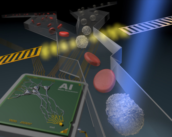
To help address these points, the physical properties of the cells can be used as an additional distinguishing feature - deformability in particular. The team headed by Jochen Guck, Director at the Max Planck Institute for the Science of Light, had developed a new technique based on this a few years ago: Real-time deformability cytometry (RT-DC). In this technique, a cell suspension is pressed through a transparent channel thinner than a hair in diameter. The cells are then stretched without damage and the degree of deformation allows an assignment to a specific cell type. The deformed cells in the channel are recorded with a high-speed camera at 2,000 to 4,000 images per second. Here, the new method of Jochen Guck and his team starts: They use a neuronal network that was trained with the images of cells marked with fluorescence markers. After the training, the artificial intelligence can identify cells without any markers and at previously unreached speed. It is even possible to sort them in real time.
This new method has many advantages: After training the neural network, the time-consuming and cost-intensive fluorescence marking for identification is no longer necessary and the cells are no longer changed by foreign molecules. At this point, the images shot by the high-speed camera are sufficient to identify the cells. This procedure is very gentle on the cells, does not change the cell properties and can analyze up to 1,000 cells per second. The application of artificial intelligence to RT-DC also offers the advantage that the parameters, on the basis of which the cell recognition or a cell change through disease can be determined, do not have to be defined beforehand. You can let the AI decide which image information is best to differentiate cells.
Guck calls the newly developed method, which has now been published in the journal Nature Methods, an "ultimate cell sorter": it combines the accuracy of the established detection via fluorescence with the sensitivity of inherent mechanical cell properties and has the potential to become a future standard method to be used in all biological and biomedical laboratories. In the future, it will be possible, for example, to quickly recover blood-forming stem cells from a sample, which can then be injected into a chemotherapy patient to rebuild the immune system, or to sort out particularly suitable photoreceptor cells from human organoids in order to avert certain forms of blindness by transplantation.
Find out more about the new cell sorter in our press release or in the original publication.
Contact
Edda Fischer
Head of Communication and Marketing
Phone: +49 (0)9131 7133 805
MPLpresse@mpl.mpg.de