
Biophotonics
Biophotonics describes the interaction of light with cells and tissues. We are interested in this interaction which is governed by the optical properties of cells. The knowledge of these optical properties is fundamental for applications developed in our group, such as the optical stretcher [1] or the optical cell rotator [2, 3] (see Optical Trapping) . The more is known about the optical properties of cells, the better become the biophysical techniques that make use of them.
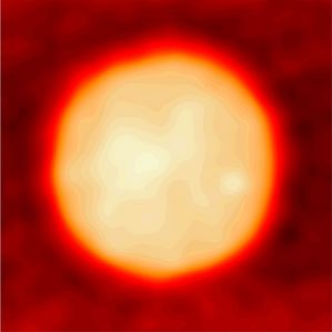
Phase image of an HL60/S4 cell recorded with digital holographic microscopy (DHM)
The optical properties of cells are defined by their internal refractive index (RI) distribution. The RI distribution generates a phase delay for light that is passing through and which can be measured using quantitative phase imaging (QPI). Note that QPI is an entirely marker-free technique, because the RI is an inherent structural property of every cell. QPI images of cells quantify their optical thickness but do not allow statements about their RI unless the shape of the cell (e.g. spherical in suspension) is known. If such structural properties are not known, optical diffraction tomography (ODT) can be used to decouple the RI from the measured optical thickness (see QPI and ODT). We have implemented several tools for QPI and ODT which we employ in our studies and which we have made available to the general public.
With our approach to biophotonics, we could show that the RI of cells is a biological marker for e.g. differentiation [4] or bacterial infection [5, 6]. In another study, we put our focus on the RI distribution within cells by studying the RI of the nucleus. The RI of the nucleus was so far generally considered higher than that of the remainder of the cell. By measuring the RI of isolated nuclei, we found that the nuclear RI is actually lower compared to the average RI of the entire cell [7]. This unexpected result has interesting and important implications for the density distribution within cells, painting a picture of a crowded cytoplasm embedding a less dense nucleus.
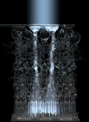
Müller cells are living optical fibers in the vertebrate retina [8].
Besides the structural information that can be extracted from the RI as a biological marker, the RI itself can play an important functional role. One peculiar example is the vertebrate retina, which is inverted with respect to its optical function. The light-sensing photoreceptor cells are located on the ‘wrong’ side — the side furthest away from the incoming light. Consequently, light has to traverse hundreds of microns of potentially scattering tissue. We have shown that there are cells in the retina that act as optical fibers and that photoreceptor cells in nocturnal animals have an inverted chromatin arrangement with a lower RI at the perimeter than at the center of the nucleus, effectively generating microlenses. Both aspects improve the light transmission through the retina and give new insight on the retina as an optical system [8, 9, 10, 11, 12, 13].
Our technical advances in biophotonics open up a new field for single-cell analysis with potential applications in medicine, including blood screening for bacterial infection or infection by the malaria parasite.
[1] J. Guck, S. Schinkinger, B. Lincoln, F. Wottawah, S. Ebert, M. Romeyke, D. Lenz, H. M. Erickson, R. Ananthakrishnan, D. Mitchell, J. Kas, S. Ulvick, and C. Bilby, “Optical deformability as an inherent cell marker for testing malignant transformation and metastatic competence,” Biophysical Journal, vol. 88, iss. 5, pp. 3689-98, 2005.
[2] M. K. Kreysing, T. Kießling, A. Fritsch, C. Dietrich, J. R. Guck, and J. A. Käs, “The optical cell rotator,” Optics Express, vol. 16, iss. 21, p. 16984, 2008.
[3] M. Kreysing, D. Ott, M. J. Schmidberger, O. Otto, M. Schürmann, E. Martín-Badosa, G. Whyte, and J. Guck, “Dynamic operation of optical fibres beyond the single-mode regime facilitates the orientation of biological cells,” Nature Communications, vol. 5, p. 5481, 2014.
[4] A. E. Ekpenyong, G. Whyte, K. Chalut, S. Pagliara, F. Lautenschläger, C. Fiddler, S. Paschke, U. F. Keyser, E. R. Chilvers, and J. Guck, “Viscoelastic properties of differentiating blood cells are fate-and function-dependent,” PLoS One, vol. 7, iss. 9, p. e45237, 2012.
[5] A. E. Ekpenyong, S. M. Man, S. Achouri, C. E. Bryant, J. Guck, and K. J. Chalut, “Bacterial infection of macrophages induces decrease in refractive index,” Journal of Biophotonics, vol. 6, iss. 5, p. 393–397, 2012.
[6] S. M. Man, A. Ekpenyong, P. Tourlomousis, S. Achouri, E. Cammarota, K. Hughes, A. Rizzo, G. Ng, J. A. Wright, P. Cicuta, J. R. Guck, and C. E. Bryant, “Actin polymerization as a key innate immune effector mechanism to controlSalmonellainfection,” Proceedings of the National Academy of Sciences, vol. 111, iss. 49, p. 17588–17593, 2014.
[7] M. Schürmann, J. Scholze, P. Müller, J. Guck, and C. J. Chan, “Cell nuclei have lower refractive index and mass density than cytoplasm,” Journal of Biophotonics, p. n/a–n/a, 2016.
[8] K. Franze, J. Grosche, S. N. Skatchkov, S. Schinkinger, C. Foja, D. Schild, O. Uckermann, K. Travis, A. Reichenbach, and J. Guck, “Müller cells are living optical fibers in the vertebrate retina,” Proceedings of the National Academy of Sciences, vol. 104, iss. 20, p. 8287–8292, 2007.
[9] I. Solovei, M. Kreysing, C. Lanctôt, S. Kösem, L. Peichl, T. Cremer, J. Guck, and B. Joffe, “Nuclear architecture of rod photoreceptor cells adapts to vision in mammalian evolution,” Cell, vol. 137, iss. 2, p. 356–368, 2009.
[10] M. Kreysing, L. Boyde, J. Guck, and K. J. Chalut, “Physical insight into light scattering by photoreceptor cell nuclei,” Optics Letters, vol. 35, iss. 15, p. 2639, 2010.
[11] M. Kreysing, R. Pusch, D. Haverkate, M. Landsberger, J. Engelmann, J. Ruiter, C. Mora-Ferrer, E. Ulbricht, J. Grosche, K. Franze, S. Streif, S. Schumacher, F. Makarov, J. Kacza, J. Guck, H. Wolburg, J. K. Bowmaker, G. von der Emde, S. Schuster, H. -J. Wagner, A. Reichenbach, and M. Francke, “Photonic crystal light collectors in fish retina improve vision in turbid water,” Science, vol. 336, iss. 6089, p. 1700–1703, 2012.
[12] Z. Blaszczak, M. Kreysing, and J. Guck, “Direct observation of light focusing by single photoreceptor cell nuclei,” Optics Express, vol. 22, iss. 9, p. 11043, 2014.
[13] M. Schürmann, G. Cojoc, S. Girardo, E. Ulbricht, J. Guck, and P. Müller, “Three-dimensional correlative single-cell imaging utilizing fluorescence and refractive index tomography,” Journal of biophotonics, vol. 11, iss. 3, p. e201700145, 2017.