

Multifocal confocal microscopy
Team Members: Gargi Sharma, Muktesh Mohan, Maria Romodina
To study the internal luminal organs such as the GI tract, a flexible endoscope with a few millimeters diameter is required. Several flexible endoscopic confocal imaging systems have been demonstrated. Unfortunately, such images are restricted to a single plane of the tissue and do not provide three-dimensional images of the tissue. Using the chromatic dispersion property of light, one may achieve a multifocal confocal microscopy system while maintaining a comparable lateral resolution. In this project, we have developed an ultralong imaging range (2 cm) chromatic confocal microscopy system with cellular-level spatial resolution. We used the exceptional chromatic dispersion properties of Zinc Selenide material to achieve the extension in the imaging range.
Polarisation-based label-free high contrast imaging
Team Members: Gargi Sharma, Muktesh Mohan, Maria Romodina, Katharina Blessing, Asha Parmar
A major research direction of our group is to develop high-contrast label-free imaging techniques. In this direction, we have developed polarisation contrast-based endoscopic optical coherence tomography (OCT) imaging techniques. Polarisation-based OCT systems exploit the birefringence and depolarisation properties of the tissue to achieve additional contrast between the different structures. Using the developed techniques, we have acquired high-contract imaging of different tissues. We believe that the simplified cross-polarised optical coherence tomography systems we have developed will be useful diagnostic tools in pathological studies.
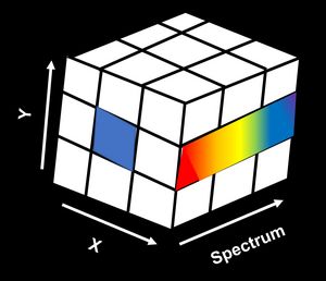
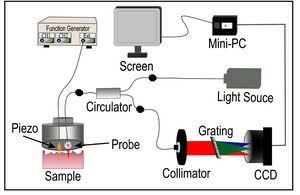
Hyperspectral imaging
Team Members: Gargi Sharma, Maria Romodina, Soumik Chatterjee
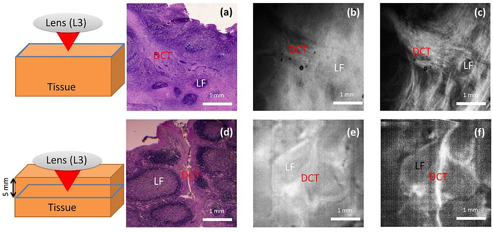
We are developing label-free hyperspectral endoscopic devices suitable for imaging GI tract in mouse models. We have combined a hyperspectral light source with a cellular resolution home-built endoscope to study the inflammatory bowel disease in a chemically induced colitis mouse model. The resolution of the endoscope is approximately 4 µm for 700 nm and approximately 8 µm for 900 nm which is sufficient to image different anatomical features such as blood vessels and colonic crypts. Since hyperspectral imaging provides spectroscopic information, this information can be used to measure the oxygen saturation of blood in vivo.
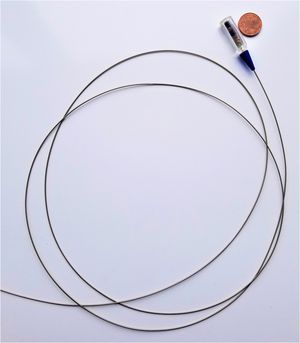
Low-cost tethered capsule for multimodal upper GI tract imaging
Team Members: Gargi Sharma, Katharina Blessing
Endoscopy is an integral part of the healthcare system. Endoscopic devices based on white light imaging, narrow band imaging and autofluorescence imaging are the most commonly used ones. Even being around for more than two centuries, endoscopy is still accessible to only a few privileged and that too with a major cost burden. In this project, we have developed a low-cost smart-phone compatible endoscopic platform for imaging the upper GI tract in low-resource settings. The smart-phone compatible endoscopic platform which is based on a low-cost USB camera can be modified to acquire images for one of the three imaging modalities, i.e. white-light imaging, narrow-band imaging, or fluorescence/autofluorescence imaging.
Skin elastography
Team Members: Asha Parmar, Maria Romodina
The goal of this project is to measure the elastic properties of skin in patients with systemic sclerosis and monitor their health over a period of time. In current clinical settings, the Rodnan skin score, based on skin palpation is still the most widely used method to determine the mechanical properties of the skin. However, the quantification and reliability of such scoring are highly dependent on the clinician’s experience. In vivo quantification of the biomechanical properties of the tissues within clinical settings has been challenging because of the unavailability of compact portable measurement devices. In this project, we have developed a portable elastography system based on optical coherence tomography.
Contact
Singh Research Group
MPI for the Science of Light
Staudtstr. 2
D-91058 Erlangen, Germany