

Real-time deformability cytometry (RT-DC) is a novel high-throughput method for the mechanical characterization of single cells that has recently been developed in our lab [3, 4, 5]. Based on the hydrodynamic deformation of cells translocating through a microfluidic channel in a contact-free manner, RT-DC is able to analyze more than 100 cells per second in real-time.
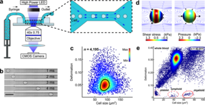
The main working principle of RT-DC is shown in the figure. Within a few milliseconds upon entry of a cell into the channel, the cell shape reaches a steady state. For each cell, several parameters can be recorded in real-time which can be visualized and gated in a post-processing step using our in-house software ShapeOut. Analytical and numerical models developed in our lab also permit the derivation of material properties such as the Young’s modulus [1, 2]. The technique is now being applied in more than 50 collaborations [6, 7, 8].
Furthermore, we have recently developed an RT-DC setup with fluorescence detection (RT-FDC). Now it is possible to not only detect the mechanical phenotype of each individual cell but also to simultaneously gather its fluorescence intensity in up to three channels in a manner similar to a conventional flow cytometer [9]. This direct correlation of mechanical with fluorescence data based on dyes, fluorescent reporter proteins or surface markers will lead to a more comprehensive validation of cell mechanics as a label-free marker.
RT-DC and RT-FDC are available as commercial products from the spin-off company ZELLMECHANIK DRESDEN GmbH.
Material available for the public domain:
1. Images of live cells obtained using real-time deformability cytometry. The cells shown originate from various tissues including lung, liver and kidney. Various physical properties, including cell deformability, are derived from every image.
https://mpl.mpg.de/fileadmin/user_upload/Guck_Division/cells_slide.png
2. A human cell passing through the microfluidic chip in a deformability cytometry instrument. The cell originally has a spherical shape and becomes deformed due to the forces acting on it upon entering the microscopic channel.
https://mpl.mpg.de/fileadmin/user_upload/Guck_Division/2a.mp4
3. Scheme of the microfluidic chip used for deformability cytometry. The cells flow through a narrow microfluidic channel, where they are deformed by shear stresses and pressure gradients. An image is taken of each cell and various physical properties are calculated from the image. Up to 1000 cells can be analyzed per second.
https://mpl.mpg.de/fileadmin/user_upload/Guck_Division/2b.png
4. Scheme of "RAPID Diagnostics", a collaborative project between the Max Planck Institute for the Physics of Light and Universitatsklinikum Erlangen, which has received the Medical Valley Award with 250 000 EUR worth of funding.
https://mpl.mpg.de/fileadmin/user_upload/Guck_Division/7.PNG
5. Standardized polyacrylamide (PAAm) beads designed as cell stiffness standards. The particles can be used as mechanical probes for the validation and calibration of cell mechanical measurements and as cell-scale stress sensors.
https://mpl.mpg.de/fileadmin/user_upload/Guck_Division/beads.png
https://mpl.mpg.de/fileadmin/user_upload/Guck_Division/beads2.png
[1] A. Mietke, O. Otto, S. Girardo, P. Rosendahl, A. Taubenberger, S. Golfier, E. Ulbricht, S. Aland, J. Guck, and E. Fischer-Friedrich, “Extracting cell stiffness from real-time deformability cytometry: theory and experiment,” Biophysical Journal, vol. 109, iss. 10, p. 2023–2036, 2015.
[2] M. Mokbel, D. Mokbel, A. Mietke, N. Träber, S. Girardo, O. Otto, J. Guck, and S. Aland, “Numerical simulation of real-time deformability cytometry to extract cell mechanical properties,” Acs biomaterials science & engineering, 2017.
[3] O. Otto, P. Rosendahl, A. Mietke, S. Golfier, C. Herold, D. Klaue, S. Girardo, S. Pagliara, A. Ekpenyong, A. Jacobi, M. Wobus, N. Töpfner, U. F. Keyser, J. Mansfeld, E. Fischer-Friedrich, and J. Guck, “Real-time deformability cytometry: on-the-fly cell mechanical phenotyping,” Nature Methods, vol. 12, iss. 3, p. 199–202, 2015.
[4] M. Herbig, M. Kräter, K. Plak, P. Müller, J. Guck, and O. Otto, “Real-time deformability cytometry: label-free functional characterization of cells,” in Flow cytometry protocols, Springer New York, 2017, p. 347–369.
[5] M. Urbanska, P. Rosendahl, M. Kräter, and J. Guck, “High-throughput single-cell mechanical phenotyping with real-time deformability cytometry,” in Methods in cell biology, Elsevier, 2018, p. 175–198.
[6] N. Toepfner, C. Herold, O. Otto, P. Rosendahl, A. Jacobi, M. Kräter, J. Stächele, L. Menschner, M. Herbig, L. Ciuffreda, L. Ranford-Cartwright, M. Grzybek, Ü. Coskun, E. Reithuber, G. Garriss, P. Mellroth, B. Henriques-Normark, N. Tregay, M. Suttorp, M. Bornhäuser, E. R. Chilvers, R. Berner, and J. Guck, “Detection of human disease conditions by single-cell morpho-rheological phenotyping of blood,” eLife, vol. 7, 2018.
[7] M. Kräter, J. Sapudom, N. Bilz, T. Pompe, J. Guck, and C. Claus, “Alterations in cell mechanics by actin cytoskeletal changes correlate with strain-specific rubella virus phenotypes for cell migration and induction of apoptosis,” Cells, vol. 7, iss. 9, p. 136, 2018.
[8] M. Urbanska, M. Winzi, K. Neumann, S. Abuhattum, P. Rosendahl, P. Müller, A. Taubenberger, K. Anastassiadis, and J. Guck, “Single-cell mechanical phenotype is an intrinsic marker of reprogramming and differentiation along the mouse neural lineage,” Development, vol. 144, iss. 23, p. 4313–4321, 2017.
[9] P. Rosendahl, K. Plak, A. Jacobi, M. Kraeter, N. Toepfner, O. Otto, C. Herold, M. Winzi, M. Herbig, Y. Ge, S. Girardo, K. Wagner, B. Baum, and J. Guck, “Real-time fluorescence and deformability cytometry,” Nature methods, vol. 15, iss. 5, p. 355–358, 2018.