

Willkommen auf der Homepage der Abteilung für biologische Optomechanik
Zellen sind die grundlegenden Einheiten biologischer Systeme. Sie haben besondere physikalische Eigenschaften, die es ihnen ermöglichen, sich in ihrer physikalischen 3D-Umgebung zu bewegen und ihre biologischen Funktionen zu erfüllen. Wir untersuchen diese physikalischen - mechanischen und optischen - Eigenschaften von lebenden Zellen und Geweben mit Hilfe neuartiger photonischer und biophysikalischer Werkzeuge, um ihre biologische Bedeutung zu testen. Unser Ziel ist der Transfer unserer Erkenntnisse in die medizinische Anwendung auf den Gebieten der verbesserten Diagnose von Krankheiten und neuer Ansätze in der regenerativen Medizin.

Von der Idee zur Anwendung: Nano Innovation Award für Nachwuchswissenschaftlerin Cornelia Holler
Das Center for NanoScience (CeNS) an der Ludwigs-Maximilian-Universität München (LMU) hat Cornelia Holler, Doktorandin am Max-Planck-Institut für die…

Physikerin Markéta Icha Kubánková mit Hermann-Neuhaus-Preis geehrt
Die Max-Planck-Gesellschaft (MPG) hat Markéta Icha Kubánková, Postdoktorandin am Max-Planck-Institut für die Physik des Lichts (MPL) und…

„Was macht ein Physiker in der Biologie und Medizin?“ – Jochen Guck hält Eröffnungsrede auf der Jahrestagung der Alexander von Humboldt-Stiftung
Am 27. und 28. Juni findet die diesjährige Jahrestagung der Alexander von Humbold-Stiftung statt. Professor Jochen Guck, Direktor am…
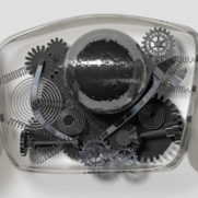
Cell Mechanics
Mechanical properties of cells are very often connected to their state and function. They can thus serve as an intrinsic biophysical marker of cell state transitions, such as metastasis of cancer cells, activation of leukocytes, or progression through the cell cycle. Read More...
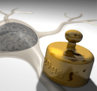
Mechanosensing
Cells actively sense and respond to a variety of mechanical signals — a process known as mechanosensing. Mechanical cues provided by the extracellular environment can modulate a wide spectrum of cellular events, including cell proliferation, differentiation and protein production. Read More...
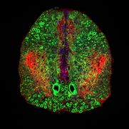
Tissue Mechanics
Cells define and largely form their surrounding tissues and, in return, receive biochemical and physical cues from them. We are working on resolving this interdependence by quantifying these tissue mechanical properties, correlating them with biological function, investigating their origin and ultimately controlling them. Read More...
Biophotonics
Biophotonics describes the interaction of light with cells and tissues. We are interested in the interaction between light and tissues which is governed by the optical properties of cells. Read More...
MPL Presseteam
Um unser Kommunikations- & Marketingteam zu kontaktieren, benutzen Sie bitte die Emailadresse: MPLpresse@mpl.mpg.de.
Weitere Informationen zu Presseanfragen oder den aktuellsten Pressemitteilungen und -bildern finden Sie auf unserer Presseseite.
Kontakt
Bitte setzen Sie sich bei Anfragen aller Art mit uns in Verbindung:
Abteilung Guck
Max-Planck-Institut für die Physik des Lichts
Staudtstr. 2
91058 Erlangen
guck-office@mpl.mpg.de
Tel: +49-9131-7133-501/-502
Fax: +49-9131-7133-990
Das Max-Planck-Institut hat seinen Sitz direkt am Südgelände der Friedrich-Alexander-Universität Erlangen-Nürnberg, auf dem die Technische Fakultät angesiedelt ist. Informationen zur Anfahrt finden Sie hier.
