Analysing tools
In order to observe the quality of produced micro and nanostructures the TDSU 1 operates different state of the art analysing tools.
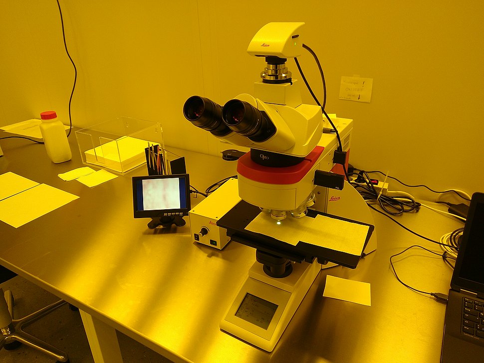
Leica DM4M
The microscope serves as non-destructive, optical investigation technique. Two DM4M optical microscopes with different objectives are available in the cleanroom.
System features:
- Dry Plan Apochromatic Objectives 2.5x ... 150x (NA 0.9), 6-position objective nosepiece
- Upright trinocular tube, eye piece 10x/25
- Koehler illumination for transmitted and reflected light, mixed illumination possible
- Yellow illumination for UV sensitive materials
- Linearly polarized illumination
- Bright field and dark field microscopy
- Full color camera system MC170HD 5MPx
- Sample stage (manual) with 4"x4" travelling range and open area
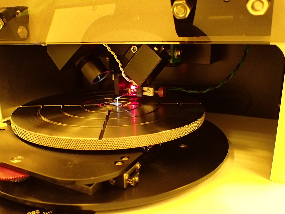
Alpha-Step D-500
A contact stylus profilometer is a tactile investigation technique for the analysis of step heights, shapes and roughness on a sample. This tool offers a 2D profiling (line scan) of the surface by moving a stylus in contact mode over the sample for a specified distance and with a specified contact force.
System features:
- Vertical range: 1200 µm, scan length: 30 mm
- Low force measurements at 0.03 to 15 mg
- Tip radius 2µm, opening angle 60°
- High resolution 5 MP color camera with 4x digital zoom for sample positioning
- Manual levelling, software levelling
- Table diameter 140mm, travelling range 20mm/80mm, 360° rotation
- Samples from a few mm size up to 4“ wafers and sample thickness up to 20 mm
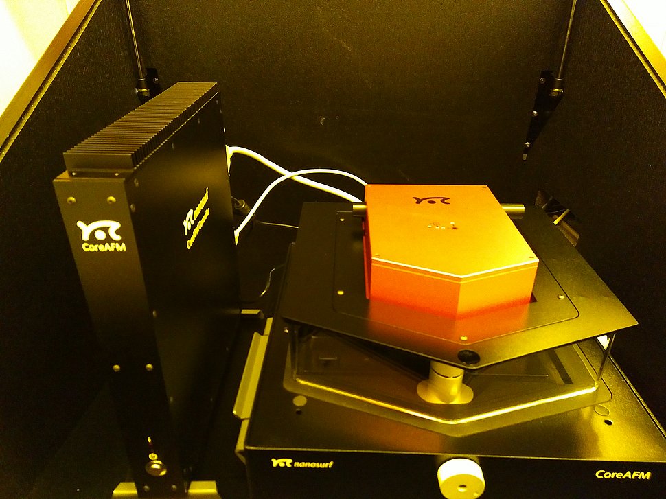
Nanosurf CoreAFM
Atomic force microscopy is especially well suited for high resolution imaging of very smooth surfaces. A cantilever with a nanometer sharp tip scans over a surface. The forces between the tip and the sample surface lead to a deflection of the cantilever. Depending on the measurement mode different information could be extracted such as surface topography or force-distance curves.
System features:
- Measurement modes: Static force microscopy, lateral force microscopy, dynamic force microscopy, spectroscopy, lithography, phase contrast microscopy
- Vertical resolution: 70 pm
- Maximum field size: 100x100 µm
- Measurable aspect ratios up to 10:1
- Sample sizes up to 50 x 50 x 5 mm
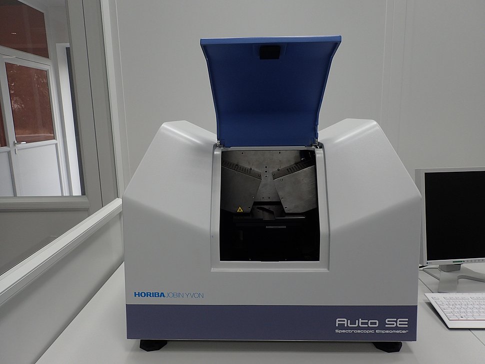
Horiba Auto SE
Ellipsometry is a non-destructive, optical investigation technique for thin film characterization. For this tool the change of polarization upon reflection is measured and compared to a model. This analysis allows the characterization of optical constants and layer thickness.
System features:
- Spectroscopic ellipsometer for the wavelength range 450nm...850nm
- Ellispometric angles Delta Δ and Psi ψ or Mueller Matrix Polarimetry (full Mueller Matrix)
- Isotropic, anisotropic and depolarizing samples, transparent samples
- Measurement spot size 25 x 60 µm ... 500 x 500 µm
- Motorized stage, sample size up to 6", sample thickness up to 5mm
- Additional camera system for sample alignment and positioning
- Elaborate software package Horiba DeltaPsi2 for advanced thin film analysis